Simultaneous determination of 7 macrolide drugs using SQ910 single quadrupole LC-MS
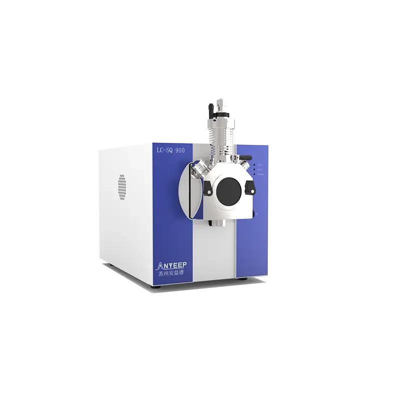
The challenge in detecting multiple macrolide residues lies not in sensitivity, but in separation and identification. Using the SQ910 single quadrupole LC-MS as an example, this article outlines the chromatographic requirements for 7 macrolides, SIM acquisition and identification boundaries, critical sample preparation points (noting erythromycin's acid sensitivity), instrument conditions, and method validation metrics aligned with standards.
Macrolide antibiotics are common targets in residue monitoring of animal-derived foods. The 7 target compounds—erythromycin, azithromycin, clarithromycin, roxithromycin, tilmicosin, tulathromycin, and josamycin—span three structural types: 14-membered, 15-membered, and 16-membered rings. Maximum residue limits (MRLs) specified in GB 31650-2019 generally range from 40 to 200 μg/kg, with erythromycin in milk having a limit as low as 40 μg/kg. Accurately analyzing all these compounds in a single injection is challenging not due to sensitivity, but because of separation and identification requirements.
Harder to detect segregation ratios
The quasi-molecular ions of 7 compounds are concentrated in the m/z range from 734 to 917. These compounds are structurally homologous and have similar polarity, leading to overlapping retention times on reverse-phase columns. Moreover, erythromycin is acid-sensitive; the presence of formic acid in the extract significantly reduces recovery rates. Sample preparation for this class of analytes must strictly control acidity rather than following generic protocols used for other veterinary drug residues.
The single quadrupole mass analyzer operates at unit mass resolution, meaning co-eluting components within the same mass window cannot be distinguished by mass alone. This makes chromatographic separation a prerequisite for method validity: gradient elution must resolve 7 peaks so that each SIM channel detects only its target analyte.
Acquisition Mode and Qualitative Boundaries
The SQ910 employs an electrospray ionization (ESI) source. In positive ion mode, macrolides generate stable [M+H]+ ions, which are collected via selected ion monitoring (SIM) on a channel-by-channel basis. The characteristic ions for the 7 target analytes are listed in the table below; however, method tuning must be re-verified on the specific instrument used.
Target | [M+H]+ (m/z, approx.) |
|---|---|
Erythromycin | 734.5 |
Azithromycin | 749.5 |
Clarithromycin | 748.5 |
Roxithromycin | 837.5 |
Tylosin | 916.5 |
Tilmicosin | 869.6 |
Josamycin | 828.5 |
Clarify the method's scope: single quadrupole provides first-stage mass filtering, with qualitative identification based on retention time and characteristic ions, and quantification via external or internal standard calibration. When regulatory requirements demand second-stage fragment confirmation, a tandem quadrupole platform must be used; this division of labor is inherent to instrument design and does not diminish SQ910's role in routine quantitative screening.
Key Control Points for Pre-processing
Animal tissue samples are extracted with acetonitrile, salted out with sodium chloride, and defatted with n-hexane. The supernatant is purified using an HLB solid-phase extraction (SPE) cartridge, dried under nitrogen to near dryness, reconstituted with mobile phase, and filtered through a 0.22 μm filter. For complex matrices such as honey and liver, matrix-matched calibration curves must be used; otherwise, matrix effects at low concentration levels will directly compromise recovery accuracy. Avoiding the introduction of formic acid during extraction is critical for ensuring stable erythromycin recovery.
Chromatography and Mass Spectrometry Conditions
Select a C18 column with dimensions 2.1 mm × 100 mm and particle size 1.7 to 1.9 μm. The mobile phase consists of 0.1% formic acid and 5 mmol/L ammonium formate in the aqueous phase, and acetonitrile in the organic phase. Perform gradient elution at a flow rate of 0.2 to 0.3 mL/min, with a column temperature of 35 to 40 °C and an injection volume of 5 to 10 μL. The total run time is 12 to 15 minutes, yielding 7 peaks for all target analytes in a single run.
Optimize the MS spray voltage, desolvation temperature, and cone voltage to ensure channel responses are within the same order of magnitude. This prevents low-response components from requiring increased sample injection, which could contaminate the ion source.
Alignment of Methodological Indicators with Standards
For the 0.5 to 200 ng/mL range, the linearity correlation coefficient for all 7 target analytes must be ≥0.995. The limit of quantification (LOQ) should be between 1 and 5 μg/kg—more than one order of magnitude below the corresponding MRL—to ensure adequate decision margins. Recovery rates at three spike levels must fall within 70% to 110%, with intra-day and inter-day relative standard deviations (RSD) <10%, which are acceptable ranges for routine monitoring. Method design may follow the cleanup and validation approaches outlined in GB/T 20762-2006, GB 31660.1-2019, and SN/T 1777.2-2007, adjusting the target analyte list based on actual matrices.